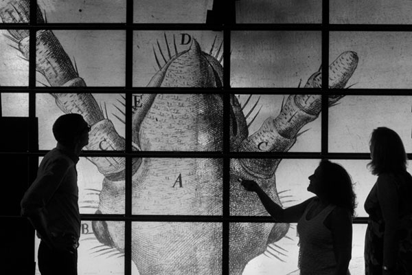
A high-resolution image of a louse captivates team members of a project that brought together the Department of English, the Texas Advanced Computing Center, and the Institute for Cell and Molecular Biology in a collaborative effort to draw attention to the Texas Institute for Literary and Textual Studies’ “The Fate of the Book” symposia by showing the essence of a physical book at the microscopic and atomic levels.
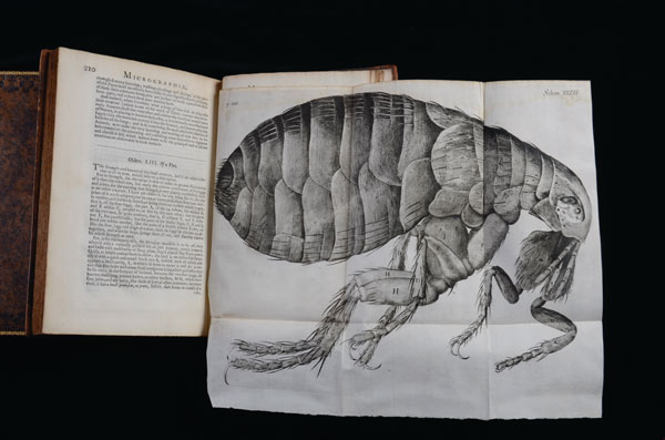
Hundreds of photos from Robert Hooke’s 1665 book, Micrographia: Or Some Physiological Descriptions of Minute Bodies, were taken with a stereo dissection microscope in the genetics lab of Janice Fischer, a professor in molecular cell and developmental biology. The photos were painstakingly shot one by one, moving the microscope’s camera across the page in millimeter increments. These photos were then stitched together into an integrated image using the expertise and processing software at the TACC/ACES Visualization Laboratory (Vislab). To view the image in its full glory, the researchers loaded it on the 36-foot-wide “Stallion” display wall at the Vislab. Finally, to get an even “deeper” perspective on the book, the team ran samples from the Micrographia’s pages through a scanning electron microscope, creating images down to the atomic level.