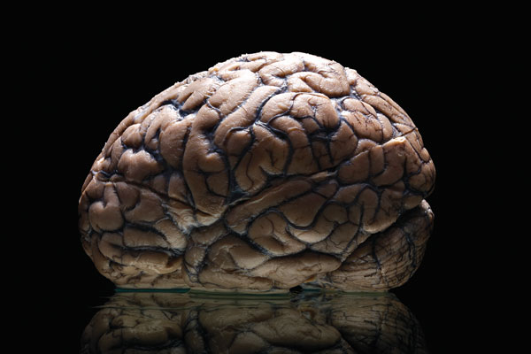
Students are busy scanning a collection of nearly 100 brains preserved from Texas State Hospital patients as part of a unique undergraduate research opportunity at The University of Texas at Austin.
A new high-resolution MRI scanner and storage space in the Norman Hackerman Building on campus makes the brain collection and associated data more accessible to students, staff and faculty.
“I think it will mean a decidedly better learning experience for students,” says psychologist Larry Cormack, who co-curates the brain collection with psychologist Tim Schallert. “When you are looking at MRI images, no matter how detailed, they have a certain level of abstraction.”
Having the actual brains as a reference will provide students with an easier way to understand the spatial relationships within the images.
College of Natural Sciences neuroscientist Jeff Luci’s class will learn to scan the brains, giving students the opportunity to learn on both the front and back end of the project.
This remarkable collection includes intact specimens dating from the 1950s to the 1980s, and includes a variety of brain abnormalities. MRI scanning leaves the preserved brains exactly as they were before, allowing them to be scanned multiple times and for long durations, a process that a living brain could not endure.
For nearly three decades, the collection remained mostly unused until now, housed in large fluid-filled jars labeled with a date of death or autopsy, a brief description in Latin and a case number—some faded with time.
“The brains have been preserved while technology has improved—similar to the way in which an archeologist might leave part of a site untouched in the hopes that better technology will come along and allow more to be learned,” Cormack says.
